

Por cortesía de Cristina Ojeda Thies (@ojedathies), Antonio Sánchez Fernández (@AnSanFer_Dr), Miguel Vázquez (@miguelvazquezdr) – voluntarios en la Iniciativa MIR 2.0 de respuesta al examen MIR de marzo de 2021. (La numeración se corresponde con la Versión 0 del examen). Gracias Dr. José Lamo de Espinosa (@lamo_espinosa) por aportar referencias para la impugnación.
Índice de contenidos
Pregunta 21:
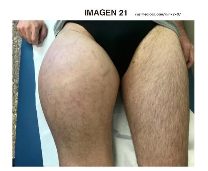
Paciente de 44 años con lesión (16x8x12 cm) de crecimiento rápido que interesa las partes blandas del tercio proximal del muslo derecho. Refiere que ha empezado a notar la masa, que tiene una consistencia dura y está adherida a planos profundos, en los últimos 6 meses. Previamente hacía deporte habitualmente, llegando a haber terminado alguna maratón. Se observa a la exploración circulación colateral, pero no se palpan adenopatías inguinales derechas. Tras la biopsia se confirma que se trata de un sarcoma pleomórfico indiferenciado de alto grado, que en los estudios de imagen se encuentra proximal al trocánter menor. La TC de extensión es negativa. ¿Cuál sería su actitud terapéutica?:
- Desarticulación de la cadera.
- Tratamiento con ifosfamida y tamoxifeno durante 6 ciclos previos a la cirugía de resección para reducir el tamaño de la lesión.
- Cirugía marginal extirpando la pseudocápsula que suelen formar estos tumores como respuesta a su rápido crecimiento.
- Hemipelvectomía derecha modificada.
Respuesta correcta: 4. Hemipelvectomía derecha modificada.
ARGUMENTO PARA LA IMPUGNACIÓN:
Pregunta a nuestro parecer IMPUGNABLE. Ninguna opción es 100% correcta. Las opciones 1 y 4 serían márgenes radicales, la 3 marginal, la 2 parece que ampliada, pero el protocolo de quimioterapia neoadyuvante no es válido.
En el enunciado de la pregunta no hay ningún dato que sugiera invasión de estructuras neurovasculares o invasión de partes blandas que impida la resección ampliada y que obligue a una cirugía radical, cosa que en cualquier caso habría que confirmar con resonancia magnética.
Es más, el tratamiento mediante cirugía radical, muy mutilante, no ha demostrado mejorar la supervivencia (hay un porcentaje importante de pacientes con micrometástasis en el momento del diagnóstico, no detectables mediante el estudio de extensión), aparte de limitar la calidad de vida. La pauta más habitual es referir al paciente a un centro especializado en sarcomas, para manejo multidisciplinar, que suele consistir en una combinación de cirugía y radioterapia, a menudo también con quimioterapia neoadyuvante, que tiene varias funciones: busca la necrosis tisular local, la respuesta al tratamiento quimioterápico ayuda en estimar el pronóstico – sobre todo si más adelante se detectan recurrencias o metástasis-, y el tratamiento sistémico puede controlar enfermedad a distancia.
La quimioterapia neoadyuvante contiene a menudo ifosfamida y doxorrubicina en 3 ciclos, mientras que el tamoxifeno (opción 2) es un agente hormonal empleado más a menudo para el cáncer de mama, y no se suelen emplear 6 ciclos, por lo cual la opción 2 también es nula.
Bibliografía:
- Marco F (Ed.) Traumatología y Ortopedia para el grado en Medicina. Elsevier, Madrid. 2015. ISBN 9788480866774. Pág 181
- Gilbert NF, Cannon CP, Lin PP, Lewis VO. Soft-tissue sarcoma. J Am Acad Orthop Surg. 2009 Jan;17(1):40-7. doi: 10.5435/00124635-200901000-00006. PMID: 19136426.
- Luis ÁM, Aguilar DP, Martín JA. Multidisciplinary management of soft tissue sarcomas. Clin Transl Oncol. 2010 Aug;12(8):543-53. doi: 10.1007/s12094-010-0552-2. PMID: 20709652.
- Cable MG, Randall RL. Extremity Soft Tissue Sarcoma: Tailoring Resection to Histologic Subtype. Surg Oncol Clin N Am. 2016 Oct;25(4):677-95. doi: 10.1016/j.soc.2016.05.014. Epub 2016 Aug 5. PMID: 27591492.
- Scoggins CR, Pisters PW. Diagnosis and management of soft tissue sarcomas. Adv Surg. 2008;42:219-28. doi: 10.1016/j.yasu.2008.04.002. PMID: 18953820.
- Grobmyer SR, Maki RG, Demetri GD, Mazumdar M, Riedel E, Brennan MF, Singer S. Neo-adjuvant chemotherapy for primary high-grade extremity soft tissue sarcoma. Ann Oncol. 2004 Nov;15(11):1667-72. doi: 10.1093/annonc/mdh431. PMID: 15520069.
Bibliografía con texto de interés.
- Marco F (Ed.) Traumatología y Ortopedia para el grado en Medicina. Elsevier, Madrid. 2015. ISBN 9788480866774. Pág 181
La cirugía de resección tumoral tiene 4 tipos de márgenes:
- Intralesional: consiste en el abordaje directo del tumor y su extirpación atravesando el mismo o, si es cavitario, vaciando su contenido mediante legrado y raspado de la pared. Esta tipo de resección solo está indicada en lesiones benignas que están limitadas por una cápsula o que son cavitarias (encondromas, lesiones quísticas, tumores de células gigantes y algunos condroblastomas).
- Marginal: a nivel de la periferia o en las proximidades del tumor y por fuera de la cápsula, justamente en la zona reactiva que la rodea. Estaría indicada en tumores benignos activos y agresivos (tumores agresivos de células gigantes, condroblastomas agresivos, osteocondromas, etc.).
- Ampliada: Incluye mínimo 1 cm de tumor sano alrededor del tumor. En realidad se trata de una resección intracompartimental y sobre todo se utiliza en tumores malignos, tanto de bajo como de alto grado, intracompartimentales.
- Radical: se realiza por fuera de los límites del compartimento, lo que implica la extirpación total del mismo junto con el tumor. Esta resección estaría indicada básicamente en el caso de tumores de alta malignidad y gran extensión local. Las amputaciones y desarticulaciones de los miembros son formas de resección radical pero muy agresivas y mutilantes, que obligan al sacrificio del miembro, por lo que están indicadas en los casos en que no lo está una cirugía de «salvación», circunstancia que se da en los siguientes casos: tumores con gran invasión de partes blandas, cuando existe infiltración de los ejes neurovasculares, en infecciones y en secuelas de la radioterapia; también se valora en los casos que se complican con una fractura patológica. Los progresos obtenidos con la cirugía conservadora han limitado mucho en la actualidad las indicaciones de la cirugía mutilante.
- Gilbert NF, Cannon CP, Lin PP, Lewis VO. Soft-tissue sarcoma. J Am Acad Orthop Surg. 2009 Jan;17(1):40-7. doi: 10.5435/00124635-200901000-00006. PMID: 19136426.
The mainstay of local treatment is excision with wide surgical margins, whenever possible. Positive margins are a risk factor for local recurrence; however, local recurrence has not been shown to affect overall survival in patients with soft-tissue sarcoma.28,29 Therefore, acceptance of close margins around vital structures is acceptable to preserve a functional limb. Limb salvage is possible in most cases, with amputation reserved for the patient in whom an adequate margin cannot be obtained with limb-sparing surgery.
Preoperative administration of chemotherapy may facilitate limb salvage by causing necrosis of the primary tumor, thereby allowing easier tumor resection. At our center, chemotherapy is usually given as a neoadjuvant treatment for highrisk tumors (American Joint Commission on Cancer stage III) or for metastatic disease (stage IV). Patients with high-grade, large, deep lesions have approximately a 50% chance of developing metastatic disease; thus, chemotherapy frequently is employed for these patients. Agents commonly used in the treatment of soft-tissue sarcoma include doxorubicin and ifosfamide.
- Luis ÁM, Aguilar DP, Martín JA. Multidisciplinary management of soft tissue sarcomas. Clin Transl Oncol. 2010 Aug;12(8):543-53. doi: 10.1007/s12094-010-0552-2. PMID: 20709652.
Amputation has been a mainstay in managing extremity STS, being used in >40% of cases [19, 20] until the early 1970s, when the hypothesis was raised that radiotherapy in combination with surgery could achieve an equivalent result [5]. Conclusions of several randomised studies confi rmed the appropriateness of limb-sparing surgery in combination with radiotherapy for a significant proportion of patients with high-grade extremity sarcomas, thus establishing a new era in their management [21]. Although it was originally thought that large high-grade sarcomas might have a lesser risk of metastasis if treated by amputation, Williard et al. reported no survival advantage for this group when treated by amputation compared with limbsparing surgery [22]… However, there remains a group of patients that is better served by an amputation rather than by a limb-sparing procedure. Consideration of amputation should be made if one or more of the following tumour characteristics occur [14] and resection of the tumour is expected to render the limb nonfunctional [3]:
1. Extensive soft tissue mass and/or skin involvement
2. Involvement of a major artery or nerve
3. Extensive bony involvement necessitating whole bone resection
4. Failure of preoperative chemotherapy or radiation therapy
5. Tumour recurrence after previous adjuvant radiation
- Cable MG, Randall RL. Extremity Soft Tissue Sarcoma: Tailoring Resection to Histologic Subtype. Surg Oncol Clin N Am. 2016 Oct;25(4):677-95. doi: 10.1016/j.soc.2016.05.014. Epub 2016 Aug 5. PMID: 27591492.
- Scoggins CR, Pisters PW. Diagnosis and management of soft tissue sarcomas. Adv Surg. 2008;42:219-28. doi: 10.1016/j.yasu.2008.04.002. PMID: 18953820.
For extremity STS, the goal of resection should be a limb‐sparing, function‐preserving oncologic resection with adequate margins… As a result, current rates of amputations for patients with STS are approximately <5% for those with primary tumors or from 9% to 14% for recurrent disease, with such procedures reserved for cases in which resection or reresection with adequate margins cannot be performed without sacrificing the functional outcome of the limb.53, 54
More recently, Gronchi et al completed a phase 3 randomized controlled trial in which high‐risk patients with high‐grade, deep, >5‐cm truncal or extremity tumors were randomized to receive standard neoadjuvant chemotherapy (anthracycline and ifosfamide) or histology‐tailored neoadjuvant chemotherapy for 5 specific sarcoma histiotypes, including UPS, myxoid liposarcoma (MLS), synovial sarcoma, malignant peripheral nerve sheath tumor, and leiomyosarcoma. With a median follow‐up duration of 12.3 months, the projected DFS rate at 46 months was 62% in the standard chemotherapy group and 38% in the histiotype‐tailored chemotherapy group (P = .004).77 After a longer follow‐up, the histiotype‐tailored chemotherapy group had better DFS than initially detected, suggesting some effect of the histiotype‐tailored chemotherapy. That trial confirms the value of neoadjuvant chemotherapy in patients with high‐risk truncal or extremity STS and further highlights the possibility of histology‐specific recruitment strategies in future randomized trials. There have also been other small retrospective series that have attempted to identify a cohort of patients with extremity STS who might benefit from neoadjuvant chemotherapy.78, 79 These studies suggest that there may be a high‐risk group of patients, such as those with high‐grade tumors measuring >10 cm, for whom neoadjuvant chemotherapy can be considered. Finally, the local impact of preoperative treatments should not be overlooked. In other words, although the primary aim of neoadjuvant chemotherapy in operable patients is systemic, a local benefit is likely to occur at least in a proportion of patients (Fig. 4). The preoperative combination of chemotherapy with radiation was shown to be feasible and to offset the adverse impact of positive surgical margins.80, 81 Function preservation may also be part of this benefit.
- Grobmyer SR, Maki RG, Demetri GD, Mazumdar M, Riedel E, Brennan MF, Singer S. Neo-adjuvant chemotherapy for primary high-grade extremity soft tissue sarcoma. Ann Oncol. 2004 Nov;15(11):1667-72. doi: 10.1093/annonc/mdh431. PMID: 15520069.
- Casali PG et al.ESMO Guidelines Committee and EURACAN. Soft tissue and visceral sarcomas: ESMO-EURACAN Clinical Practice Guidelines for diagnosis, treatment and follow-up. Ann Oncol. 2018 Oct 1;29(Suppl 4):iv51-iv67. doi: 10.1093/annonc/mdy096
Pregunta 107:
En la patología de la mano, ¿cuál de las siguientes afirmaciones sobre la enfermedad de Dupuytren es INCORRECTA?:
- Afecta con mayor frecuencia a los dedos anular y meñique.
- El tratamiento conservador con fisioterapia es poco eficaz.
- Es un engrosamiento y retracción de la aponeurosis palmar.
- Es más frecuente en trabajadores manuales.
Respuesta correcta: 4. Es más frecuente en trabajadores manuales
ARGUMENTO PARA LA IMPUGNACIÓN:
Pregunta a nuestro parecer IMPUGNABLE, ya que todas las opciones dadas son correctas. Consideramos que con la bibliografía actual puede defenderse que el enunciado 4 también es correcto.
Razonamiento: la Enfermedad de Dupuytren afecta por orden de frecuencia los dedos 4º -> 5º -> 3º -> 2º o 1º. Es un una metaplasia de las células de la aponeurosis palmar que da lugar al engrosamiento y la retracción de la misma. Se ha descrito como factor de riesgo, entre otros, los traumatismos repetidos. La asociación con la ocupación es un tema de bastante debate – si se acepta que es más frecuente en trabajadores expuestos a vibraciones, y varios trabajos recientes sí que la reconocen como enfermedad ocupacional, con un odds ratio de al menos 2. La fisioterapia es poco eficaz; aunque el tratamiento con ortesis, infiltraciones y ondas de choque puede retrasar la progresión de la enfermedad en fases precoces, pero no han demostrado la eficacia de la cirugía.
Bibliografía:
- Marco F (Ed.) Traumatología y Ortopedia para el grado en Medicina. Elsevier, Madrid. 2015. ISBN 9788480866774 : Pág 254:
- Green’s Cirugía de la mano. Capítulo 5 Contractura de Dupuytren Autor: D.A. McGrouther Pg 161
- Lurati AR. Dupuytren’s Contracture. Workplace Health Saf. 2017 Mar;65(3):96-99. doi: 10.1177/2165079916680215. Epub 2017 Jan 9. PMID: 28068478.
- Descatha A, Jauffret P, Chastang JF, Roquelaure Y, Leclerc A. Should we consider Dupuytren’s contracture as work-related? A review and meta-analysis of an old debate. BMC Musculoskelet Disord. 2011 May 16;12:96. doi: 10.1186/1471-2474-12-96. PMID: 21575231; PMCID: PMC3123614.
- Liss GM, Stock SR. Can Dupuytren’s contracture be work-related?: review of the evidence. Am J Ind Med. 1996 May;29(5):521-32. doi: 10.1002/(SICI)1097-0274. PMID: 8732927.
- Alser OH, Kuo RYL, Furniss D. Nongenetic Factors Associated with Dupuytren’s Disease: A Systematic Review. Plast Reconstr Surg. 2020 Oct;146(4):799-807. doi: 10.1097/PRS.0000000000007146. PMID: 32970002.ç
- Descatha A, Bodin J, Ha C, Goubault P, Lebreton M, Chastang JF, Imbernon E, Leclerc A, Goldberg M, Roquelaure Y. Heavy manual work, exposure to vibration and Dupuytren’s disease? Results of a surveillance program for musculoskeletal disorders. Occup Environ Med. 2012 Apr;69(4):296-9. doi: 10.1136/oemed-2011-100319. Epub 2012 Jan 2. PMID: 22213840; PMCID: PMC3815440.
Bibliografía con texto de interés.
- Marco F (Ed.) Traumatología y Ortopedia para el grado en Medicina. Elsevier, Madrid. 2015. ISBN 9788480866774 :
Pág 254: “ENFERMEDAD DE DUPUYTREN:
Esta enfermedad consiste en un engrosamiento nodular con acortamiento de la aponeurosis o fascia palmar. Es idiopática pero se ha descrito un comportamiento genético dominante, que se da especialmente en personas del norte de Europa. Afecta sobre todo a varones mayores de 50 años y se han asociado como factores de riesgo la epilepsia, la diabetes, la enfermedad pulmonar crónica, el alcoholismo, el tabaquismo y los microtraumatismos de repetición.
… El dedo más frecuentemente afectado es el cuarto, seguido, por orden de frecuencia, del quinto, del tercero, del segundo y del primero.
… El empleo de férulas no es curativo, aunque puede retrasar la progresión de las contracturas. El tratamiento definitivo es quirúrgico… “
- Green’s Cirugía de la mano. Capítulo 5 Contractura de Dupuytren Autor: D.A. McGrouther Pg 161
El mismo Dupuytren relacionó la contractura con trabajos pesados en un paciente, un chófer, y con un traumatismo en otro, un comerciante de vinos, que había trabajado hacía muchos años a un ritmo muy fuerte levantando barriles. Ninguno de estos dos tipos de casos, en que se presenta el uso derivado de la ocupación, o el traumatismo como causas, han sido sometidos a un escrutinio científico concienzudo
- Lurati AR. Dupuytren’s Contracture. Workplace Health Saf. 2017 Mar;65(3):96-99. doi: 10.1177/2165079916680215. Epub 2017 Jan 9. PMID: 28068478.
- Descatha A, Jauffret P, Chastang JF, Roquelaure Y, Leclerc A. Should we consider Dupuytren’s contracture as work-related? A review and meta-analysis of an old debate. BMC Musculoskelet Disord. 2011 May 16;12:96. doi: 10.1186/1471-2474-12-96. PMID: 21575231; PMCID: PMC3123614.
The meta-OR for manual work was 2.02[1.57;2.60] (HQMC studies only: 2.01[1.51;2.66]), and the meta-OR for vibration exposure was 2.88 [1.36;6.07] (HQMC studies only: 2.14[1.59;2.88]).
- Liss GM, Stock SR. Can Dupuytren’s contracture be work-related?: review of the evidence. Am J Ind Med. 1996 May;29(5):521-32. doi: 10.1002/(SICI)1097-0274. PMID: 8732927.
Bennett [1982: Br J Ind Med 39:98-100] found the prevalence of DC at a British PVC bagging and packing plant in which workers were exposed to repetitive manual work to be 5.5 times that at a local plant without packing, and twice the expected prevalence in a U.K. working population previously studied by Early [1962: J Bone Joint Surg 44B:602-613]. DC was observed more frequently among vibration white finger claimants than controls by Thomas and Clarke [1992: J Soc Occup Med 42:155-158] (OR, 2.1; 95% CI, 1.1-3-9), and more frequently among vibration-exposed workers than controls by Bovenzi et al. [1994: Occup Environ Med 51:603-611] (OR, 2.6 95% CI, 1.2-5.5). Cocco et al [1987: Med Lav 78:386-392] found that a history of vibration exposure occurred more frequently among cases of DC than among controls (OR, 2.3; 95% CI, 1.5-4.4). The latter two studies presented some evidence of a dose-response relationship. There is good support for an association between vibration exposure and DC.
- Alser OH, Kuo RYL, Furniss D. Nongenetic Factors Associated with Dupuytren’s Disease: A Systematic Review. Plast Reconstr Surg. 2020 Oct;146(4):799-807. doi: 10.1097/PRS.0000000000007146. PMID: 32970002.ç
There was strong evidence for the association between Dupuytren’s disease and advanced age, male sex, family history of Dupuytren’s disease, and diabetes mellitus. Furthermore, heavy alcohol drinking, cigarette smoking, and manual work exposure showed a significant dose-response relationship.
- Descatha A, Bodin J, Ha C, Goubault P, Lebreton M, Chastang JF, Imbernon E, Leclerc A, Goldberg M, Roquelaure Y. Heavy manual work, exposure to vibration and Dupuytren’s disease? Results of a surveillance program for musculoskeletal disorders. Occup Environ Med. 2012 Apr;69(4):296-9. doi: 10.1136/oemed-2011-100319. Epub 2012 Jan 2. PMID: 22213840; PMCID: PMC3815440.
Heavy manual work without vibration exposure was significantly associated with the condition (adjusted OR (aOR) 3.9; 95% CI 1.3 to 11.5) adjusted on age and diabetes), as was use of vibrating tools (aOR 5.1; 2.1 to 12.2). These associations remained significant among subjects with >10 years in the same job, with increases in aOR of 6.1 (1.5 to 25.0) and 10.7 (3.4 to 34.6), respectively.
Puedes comentar sobre este tema en el foro
OS PEDIMOS DESDE AQUÍ LA MÁXIMA DIFUSIÓN A LA INICIATIVA PARA QUE LLEGUE AL MÁXIMO DE OPOSITORES MIR PRESENTES Y FUTUROS PARA QUE LES SEA DE AYUDA.


